Monday, October 31, 2016

Is troponin really becoming a lousy test?

Just a quick reminder this week regarding the use of troponin. This is one of the most abused tests in Emergency Departments these days.  The hs Troponins seems to be overtly sensitive that might lead to unnecessary admissions. Here are a few caveats with troponin:

1. Do a history first, followed by a physical exam and then ECG. Only after that, send a troponin if you are concerned about the possibility of ACS. 

DO NOT ORDER TROPONIN FOR EVERYONE WALKING IN WITH CHEST PAIN

2. Remember that there a  plenty of other reasons to have a positive troponin besides ACS. To name a few - PE, Heart Failure, Sepsis, Renal Failure, Stroke, Tachycardia, Infiltrative Heart Disease, Myopericarditis.

A RAISED TROPONIN DOES NOT ALWAYS EQUAL ACS. TROPONIN IS A MARKER OF ANY MYOCARDIAL INJURY, NOT JUST ISCHEMIA. 

3. STEMI does not need a troponin. Focus on rapid Reperfusion (Thrombolytics or PCI). Troponin is done to differentiate between NSTEMI and USA (Unstable Angina).


4. Discharging someone with a single negative troponin is high risk business (unless it was drawn 6 hours after the onset of symptoms). Whenever possible, get two serial troponins to see if there is any significant rise form the baseline. However, serial sampling of hs troponin at 0 and 2 hours can safely rule-out of STEMI and NSTEMI. 

In case, you happen to discharge home a patient with two negative troponins, document why are you doing that because it can still be Unstable Angina. 

5. Troponin might take 6-12 hours to rise from the onset of symptoms and stays elevated upto 2 weeks. Document your concerns while sending these cardiac biomarkers.


Attorneys focus on the documentation of HPI, documentation of ECG findings and Medical Decision Making with appropriate reasoning before settling the case. We are going to cover these bits next week. 


BOTTOMLINE: I think troponin is still a great test, if done with caveats. However, over the last two decades, it has become extremely sensitive and thus non-specific (like d-dimer for PE). 


Further Reading: 


Author:

              
     Lakshay Chanana
     
     Speciality Doctor
     Northwick Park Hospital
     Department of Emergency Medicine
     England

     @EMDidactic


Monday, October 24, 2016

Hundred thousand views

A big thanks to each and everyone of you out there who supported the existence of this blog. To be honest, being the first one from India was quite intimidating but I am pleased to know that the blog has recently crossed the milestone of 100,000 hits in less than 2 years. Things were pretty rough when I began this endeavour in January 2015. There were critics and skeptics who questioned every bit of it. Fortunately, we have come a long way since then and now more and more people are open to Free Open Access Medical Education.


A special mention about all the contributors for taking out time from their busy schedules. It was not possible to reach this far without your support. Thanks a ton guys!!

  • Akshay Kumar
  • Apoorva Chandra
  • Azharullah Khan
  • Kritika Atrey 
  • Liaquat Roopesh
  • Nikhil Tambe
  • Sagar Galwankar

I usually post a topic on every Monday and it would not have been possible without the support of Karishma, my wife. She has sacrificed numerous weekends and whenever it was about the blog - there were no questions asked! She has been a huge help and also one of the major critics of this blog. 


Over the course of last couple of years, many folks asked me about the blog logo. Here is what it signifies..


The logo took its inspiration from Emergency Medical Services. It is comprised of the star of life - a symbol most commonly associated with EMS. The six branches of the star symbolise 
  1. Early Detection
  2. Early Reporting
  3. Early Response
  4. On Scene Care
  5. Care in Transit
  6. Transfer to Definitive care
The snake emblem is the Rod of Asclepius is the symbol of medical care, named for the Greek mythological figure Asclepius, who was said to have possessed healing power.

The numerous yellow colored light bulbs within and around the star represent dandelions of ideas. The ideas do have their origin and base within Emergency Medicine but they travel out to other zones to disperse and germinate as novel thoughts and innovations. 

Credits to Kritika Atrey for crafting this logo for the blog. 


And for those of you who read and COMMENT on the posts, just to let you know that you  people make my day. A word of appreciation means a lot and keeps me going. Your intrigue questions, thoughts and comments always open an opportunity for discussions.


Lastly, this note is incomplete without acknowledging the contribution of "The Teaching Course" which played an indispensable role behind the inception of this blog. This course was phenomenal and it turned out to be a life-changing experience for me. It was absolutely worth travelling over 8000miles to be there. I owe it to these people.


Click here to know more about the teaching course. 


Author:


     Lakshay Chanana
     
     Speciality Doctor
     Northwick Park Hospital
     Department of Emergency Medicine
     England

     @EMDidactic


Monday, October 17, 2016

Hyperlactatemia and Lactic Acidosis

I recently came across a young patient with unexplained hyperlactatemia. He came in with severe abdominal pain and was found to have a normal CT Abdomen with a background of ethanol related chronic pancreatitis . He was eventually admitted for pain relief and unexplained hyperlactatemia. So I went back and did a bit of reading on LACTATE...
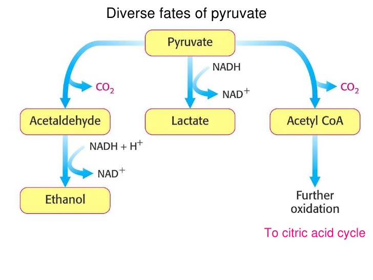
What is Lactate?
Lactate is the normal endpoint of the anaerobic breakdown of glucose. Most of the lactate production occurs in skeletal muscle, bowel, brain, and RBCs. The lactate generated can be taken up by the liver and converted to glucose (via gluconeogenesis) or can be used as a primary oxidative fuel. In the setting of decreased tissue oxygenation, lactic acid is produced as the anaerobic cycle is utilized for energy production.


Evidence suggests increased morbidity and mortality for patients with  increasing lactate levels or a decreased rate of lactate clearance.


How is lactate cleared from our body?
Lactate is cleared from blood by thru liver, kidneys (10-20%) and skeletal muscles. The ability of the liver to consume lactate is concentration-dependent and progressively decreases as the level of blood lactate increases. 
Lactate producers: skeletal muscle, the brain, the gut, and the erythrocytes. 
Lactate metabolizers: Liver, the kidneys, and the heart.


Lactate v/s Lactic Acid and Hyperlactatemia v/s Lactic Acidosis.
Lactate is not synonymous with lactic acid, and hyperlactatemia is not synonymous with lactic acidosis.
Hyperlactatemia is defined as a persistent, mild to moderate (upto 4-5 mmol/L) increase in blood lactate concentration without metabolic acidosis. It can occur in the setting of adequate tissue perfusion, intact buffering systems, and adequate tissue oxygenation.

Lactic acidosis is characterized by persistently increased blood lactate levels (usually >5 mmol/L) in association with metabolic acidosis. Also, lactic acidosis may not necessarily produce acidemia in a patient. The development of lactic acidosis depends on the magnitude of hyperlactatemia, the buffering capacity of the body, and the coexistence of other conditions that produce tachypnea and alkalosis (eg, liver disease, sepsis). Thus, hyperlactatemia or lactic acidosis may be associated with acidemia, a normal pH, or alkalemia.

D-Lactate and L-lactate

L-lactate and D-lactate are the two isomeric forms of lactate.
L-lactate is the most commonly measured level, as it is the only form produced in human metabolism.

D-lactate is a byproduct of bacterial metabolism and may accumulate in patients with short-gut syndrome or in those with a history of gastric bypass or small-bowel resection

What causes an elevated lactate?
  1.   Tissue Hypoxia and Anaerobic Metabolism (Traditional school of thought)
  2.   Due to decreased clearance rather than increased production in sepsis
  3.   Secondary to down-regulating of pyruvate dehydrogenase in skeletal muscles by inflammatory mediators (Cytokines) and Catecholamines.


Is it possible to have hypoperfusion but a normal lactate level?
Short Answer- YES
For significant increase in blood lactate to occur, lactate must be released into the systemic circulation and the rate of production must exceed hepatic, renal, and skeletal muscle uptake. Therefore, regional hypoperfusion of tissues may be present despite normal blood lactate concentrations.

Types of Lactic Acidosis
  • Type A – Poor tissue perfusion or oxygenation of blood (eg, hypotension, cyanosis, cool and mottled extremities). It can be caused by the overproduction of lactate or the underutilization of lactate. For instance, muscular activity, seizures, ischemia, shock, hypoxemia, anemia, CO poisoning.

  • Type B - no clinical evidence of poor tissue perfusion or oxygenation exists.
  1. Type B1 occurs in association with systemic disease, such as renal and hepatic failure, diabetes and malignancy, thiamine deficiency, infection, pancreatitis, short bowel syndrome
  2. Type B2 is caused by several classes of drugs and toxins, including biguanides, alcohols, iron, isoniazid, zidovudine, and salicylates.
  3. Type B3 is due to inborn errors of metabolism.

List of Possible Causes
  •       Sepsis related Hypoperfusion and Mitochondrial Dysfunction
  •       Bowel ischemia
  •       Severe iron-deficiency anemia, Diabetes mellitus
  •       Liver disease, Kidney Disease
  •       Alcoholic Ketoacidosis, Pancreatitis
  •       Malignancy (leukemia, lymphoma, lung cancer)
  •       Seizures
  •       Heat stroke, Pheochromocytoma
  •       Thiamine deficiency (Remember this one during ICU rounds!)
  •       Inborn errors of metabolism - von Gierke disease, fructose-1,6-diphosphatase  deficiency, pyruvate carboxylase deficiency, pyruvate dehydrogenase deficiency, oxidative phosphorylation deficiency, and methylmalonic aciduria.
  •       MELAS syndrome (mitochondrial encephalopathy, lactic acidosis, and stroke like episodes) - Characterized by migraine like headaches, dementia, hearing loss, ataxia, and episodic vomiting


What drugs can cause an elevated lactate?
   Acetaminophen
   Alcohols and glycols (ethanol, ethylene glycol, methanol, propylene glycol)
   Antiretroviral nucleoside analogs (zidovudine, didanosine, lamivudine)
   Beta-adrenergic agents (epinephrine, ritodrine, terbutaline, salbutamol)
   Biguanides (phenformin, metformin)
   Cocaine
   Cyanogenic compounds (cyanide, nitroprusside)
   5-FU
   Iron, Isoniazid
   Propofol
   Salicylates
   Sulfasalazine
   Valproic acid


Click here to listen to Scott Weingart talking about Lactate

Take Home:

  • Get familiar with Type B causes of an elevated lactate
  • See the medication list and co-morbities while evaluating elevated lactate


Other Resources:
EMCrit/Pulmcrit on Lactate
Resus.me 


Author:

              
     Lakshay Chanana
     
     Speciality Doctor
     Northwick Park Hospital
     Department of Emergency Medicine
     England

     @EMDidactic


Monday, October 10, 2016

The Acutely Confused Elderly Patient


Patients presenting to the ED with an Altered Mental Status (AMS) require a thorough history and an extensive work-up leading to time consuming evaluation, talking with families and caregivers, reviewing old records, labs and radiologic studies. Even after an broad work up, occasionally a diagnosis is not reached. It becomes a challenge for emergency physicians to work with limited history and examination findings. Therefore, adopting an organized approach to the evaluation of mental status would result in increased clinician comfort while taking care of elderly, better ability to communicate with other physicians, and improved patient and family satisfaction. 

Why do we need to learn about this?
ED visits for elderly patients are predicted to increase in future and at least 1/4 of all ED patients over age 65 years have some form of AMS. As the first responders, we need to be comfortable while evaluating the patients. 

What causes this acute confusional state?
A normal level of consciousness depends on an intact reticular activating system, cerebral cortex, and communication between the two. Various pathologies may disrupt optimal cortical functioning and result in confusion and the pathophysiology is not straightforward to understand. Widespread cortical dysfunction may result from substrate deficit, neurotransmitter dysfunction, or circulatory dysfunction. The magnitude of problem also depends on the reserve of central nervous system (CNS) function which varies from individual to individual; people with a pre-existing impairment may become confused after even minor changes in their normal state.


Delirium 
Delirium is an acute, fluctuating change in cognition, accompanied by impaired attention and consciousness. At times patient may be awake and distracted, then progress to a hyperalert state, and later may be lethargic. The mortality associated with delirium changes depending on whether or not the diagnosis is made in the ED (or in hospital). Although many patients recover fully, but they are prone have an increased likelihood of persistent cognitive deficits. 

Diagnosis of delirium
Delirium can present with:
  1. Hypoactive i.e. lethargic
  2. Hyperactive i.e. hyperalert
  3. Mixed i.e. alterations between hyperactive and hypoactive.
  4. Normal psychomotor activity
Delirium occurs abruptly and is usually apparent within hours to days of onset. Patients may have a prodrome of restlessness, impaired attention, and sleep disturbance that can last several days before the onset of frank delirium. It is this criterion that differentiates delirium from dementia.

Other symptoms of delirium 
  • Altered sleep–wake patterns (daytime drowsiness and nighttime agitation and disorientation i.e. sundowning)
  • Nonfocal neurologic deficits may occur, such as speech and language deficits (dysarthria, dysnomia, dysgraphia, or aphasia).
  • Asterixis is associated with hepatic, renal disease or hypercarbia.
  • Nystagmus and cerebellar abnormalities may suggest alcohol or drug intoxication.
  • Pupillary abnormalities also can suggest drug intoxication (eg, miosis with narcotics).
  • Alcohol or sedative–hypnotic withdrawal is associated with coarse tremors, tachycardia, and low-grade fever.
  • Anticholinergic toxidrome (dry mouth, urinary retention, tachycardia, fever).

Differentiating Delirium from Dementia
Both delirium and dementia may cause impaired cognition. The key is finding out the onset of the symptoms; in delirium the symptoms are acute, whereas in dementia the onset is longer and more subtle. Determination of a single etiology causing delirium may be difficult, and often more than one etiology contributes to the delirium. Also remember that demented patients may presented with acute delirium with a long standing underlying dementia. 
Another possible cause for patients presenting with delirium includes primary psychiatric disorders, such as acute psychosis. This diagnosis, however, should be made only in patients without a prior history of psychiatric disease after extensive evaluation, rather than in the ED.

Etiology
Delirium is a manifestation of various medical disorders of cerebral metabolism or neurotransmission, and as such the etiologies are broad. 
1. Primary intracranial disease

  • Cerebrovascular Accident

  • Acute or Chronic Subdural hepatoma 
  • Encephalitis
/ Meningitis

  • Seizures (Convulsive or Non-Convulsive)
  • Postictal state 
  • Neoplasm
  • Raised ICP
2. Systemic diseases secondarily affecting the central nervous system
  • Hypertensive encephalopathy
  • Infections (Pneumonia
/ UTIs/ Skin and soft tissue infections)
  • Cardiopulmonary disorders (Acute myocardial infarction, CCF, Arrhythmia, Cardiogenic shock, 
Acute or chronic respiratory failure)
  • Uremia
  • Hepatic encephalopathy

  • Fluid or electrolyte abnormalities (Dehydration. Hyper/HypoNa, Hypo/Hyperglycemia
, Hyper/HypoCa)
  • Nutritional (Severe Anemia)
  • Thyroid/Adrenal Disorders
  • Paraneoplastic Syndromes
3. Exogenous toxins and Drug withdrawal
  • Anticholinergic medications - Antihistamines, 
Antiemetics, 
Antiparkinsonian medications 
  • Antispasmodics (gastrointestinal) Alcohol
  • Sedative–hypnotics

Common etiologies in elderly patients include infections, medications, and primary CNS disorders. Focus of an infection can be urine, respiratory tract, intrabdominal pathology, skin and soft tissues or CNS. If they look unwell, administer broad spectrum antibiotics and obtain relevant cultures. 
Don’t miss on the drug history including over the counter medications. The most common category of medications to cause delirium is drugs with anticholinergic properties: antihistamines, antiemetics, antipsychotics, antiparkinsonian drugs, antidepressants, gastrointestinal antispasmodics and fentanyl patches. It is imperative to review the side-effect profile of any new medication and to assume that a new medication as culprit if alternative explanations are not available.

What kind of work up do they need in the ED?
The workup of an older patient who has delirium can be extensive unless a clear cause is determined from the history and physical examination. This may be difficult and frustrating, if a history from a surrogate is not available and the patient is unable to cooperate with the examination. The mortality associated with delirium is high, especially if unrecognized. 
  • CBC (infections are a common cause of delirium in elderly and they may present without fever or leukocytosis!
  • Electrolytes (Hypo/HyperNa, Hyper/HypoCa)
  • BUN, Creatinine (Uremia)
  • Glucose (Hypoglycemia)
  • EKG (ACS may present with confusion in elderly)
  • Pulse Oximetry (Hypoxia)
  • ABG (Hypercarbia, Hypoxia)
  • CXR (Pneumonia, CHF)
  • UA (UTI)
  • CT Head/LP (Stroke, Meningitis)
  • Serum Ammonia (if they have a h/o liver issues)
  • Urine Dug Screen
  • Thyroid Function
  • Bedside USG (Look for urinary retention, cardiac function, consolidation)

The yield use of computed tomography (CT) of the brain in patients who have delirium is not great unless you have focal new neurological findings.

Role of Family and Caregivers
It is pivotal to understand the family’s position and perspective here, as they are the ones who take care of the patient. Avoid medical jargon when explaining the prognosis, be realistic about the outcome and arrange support for them if needed. Also get a sense of a patient’s baseline status before you begin your evaluation. Live-in family may be able to provide a clearer history of the duration of symptoms than others who haven’t seen the patient for long. In nursing home patients, it is helpful to contact the nurse who sent the patient to fill in the gaps in the history.
Determinations of the level of consciousness can be made by simple observation of the patient during routine history and examination. A thorough history including, dietary habits, symptoms of thyroid disease, alcohol, medications etc. goes a long way.

Emergency Management of an Acutely Confused Patient
Treatment of delirium usually is directed at the underlying cause of the delirium. Identify and treat the life threatening causes such as STEMI, Seizures, Hypoxia quickly. If a cause is not identified in the ED, then admit them and do the additional work up. Minimise environmental interventions, such as turning off the lights, bringing families to the bed-side. Avoid physical restraints if possible as they increase the patient’s agitation and the severity of the delirium. 
They might require some fluids, antibiotics and other medications to allay the agitation. 
Haloperidol is recommended most frequently in low doses i.e. 0.5 to 1.0 mg orally, IM, or IV to control the agitation (Repeat every 30 minutes until the desired effect is achieved). Other options include risperidone or olanzapine. Use benzodiazepines only for specific conditions, like alcohol or sedative hypnotic withdrawal or seizures. Beware of paradoxic CNS reactions that may worsen the patient’s condition.

Disposition
Older ED patients should be admitted to the hospital for evaluation unless there is a single, clear, and reversible etiology of the delirium, such as intoxication from a short-acting medication. 

Take Home:

  • When evaluating delirium, do a through history and physical and ask for medications and also over the counter prescriptions.
  • Identify and treat life threats first.
  • Do not underestimate the role of family or nursing home staff who can better detect a change in the patient’s cognition. Be extremely sensitive while dealing with the family.

Author:

              
     Lakshay Chanana
     
     Speciality Doctor
     Northwick Park Hospital
     Department of Emergency Medicine
     England

     @EMDidactic